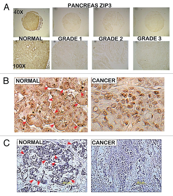
Figure 2.
ZIP3 immunohistochemistry of tissue array slides and tissue sections containing normal pancreas vs. adenocarcinoma cores. The same tissue array series as employed in Figure 1. (A) Panels show ZIP3 IHC for normal and adenocarcinoma cores. (B) High magnification shows basal membrane localization (arrows) of ZIP3 in normal epithelium and absence of ZIP3 transporter in adenocarcinoma. (C) ZIP3 IHC of tissue sections of normal pancreas tissue vs. adenocarcinoma provided and performed by Dr. Desouki. Confirms the localization of ZIP3 with basal membrane of epithelium and the absence of ZIP3 in adenocarcinoma.

