Figure 1.
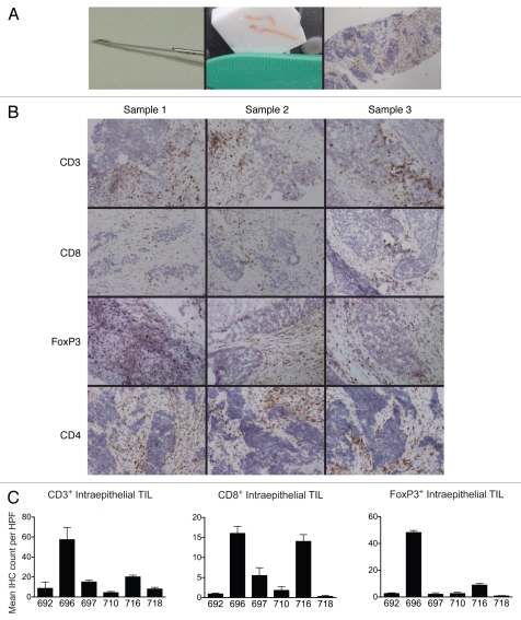
(A) Tumor biopsy acquisition and histology. Left, 16-gauge Quick-Core* biopsy needle. Center, tumor specimen obtained with two needle passes, embedded in O.C.T. Right, representative immunohistochemical section, anti-CD3-DAB stain, hematoxylin counterstain, 40× original magnification. (B) Representative immunostains of triplicate specimens; anti-CD3, CD8, FoxP3 and CD4 stains, 40× original magnification. (C) Mean intratumoral CD3+, CD8+ and FoxP3+ TILs for each patient, reflecting the mean of 5, 200× high-power fields. Error bars indicate standard deviation of the mean.

