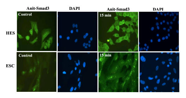
Figure 4.
Immunofluorescence localization of Smad3 in HES and ESC. The cells were incubated under serum-free condition for 24 hrs then treated with GnRH (0.1 μM) for 5, 15 and 30 min. Note subcellular localization of Smad3 in untreated control with mostly cytoplasmic and limited nuclear localization, while GnRH-treatment resulted in more cytoplasmic accumulation of Smad3. The figures are shown after 15 min of GnRH treatment with FITC staining used to localize Smad3 and DAPI staining for the nuclei.

