Figure 5.
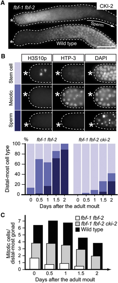
Ectopic expression of CKI-2 results in stem cell loss in FBF(−) animals. (A) CKI-2 is expressed in distal-most gonads of fbf-1 fbf-2 animals. Immunodetection of CKI-2 in distal gonads (outlined), dissected at 0.5 days after the adult moult, from animals of the indicated genotypes. The proximal fbf-1 fbf-2 gonad (arrow) contains sperm and therefore does not express CKI-2. (B) Stem cell loss in fbf-1 fbf-2 mutants is largely rescued by removing cki-2. Top: Distal-most cells of fbf-1 fbf-2 mutant gonads dissected at 1 day after the adult moult and stained for the indicated markers. Stem cells do not express the meiotic marker HTP-3, and those in mitosis are positive for H3S10p staining which marks condensed chromosomes. Conversely, loss of H3S10p staining and expression of HTP3 indicates meiotic entry. Nuclei of germ cells that differentiated into sperm have a characteristic dot-like appearance visualized by DAPI staining. Bottom: Fractions of gonads that in the distal-most part contain: stem cells (light blue), meiotic cells (intermediate blue), or sperm (dark blue), measured at 12 h intervals after the larval-to-adult moult. (C) Depletion of CKI-2 from fbf-1 fbf-2 gonads partially restores stem cell proliferation. Numbers of H3S10p-positive (mitotic) cells in distal-most gonads from animals of indicated genotypes are shown. Number of gonads analysed for panels (B) and (C) fbf-1 fbf-2/fbf-1 fbf-2 cki-2/wild type: d0=58/45/44, d0.5=43/28/48, d1=41/43/13, d1.5=7/34/31, d2=43/19/44.

