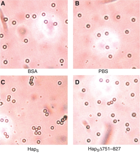
Figure 7.
Visualization of HapS-coated beads. Latex beads were coated with either BSA (A), HapS (C), or HapSΔ751–827 (D) and were viewed by light microscopy. Beads coated with HapS formed aggregates, while beads coated with HapSΔ751–827 remained isolated, similar to controls (BSA-coated beads and beads incubated in PBS alone (B)).

