Figure 2.

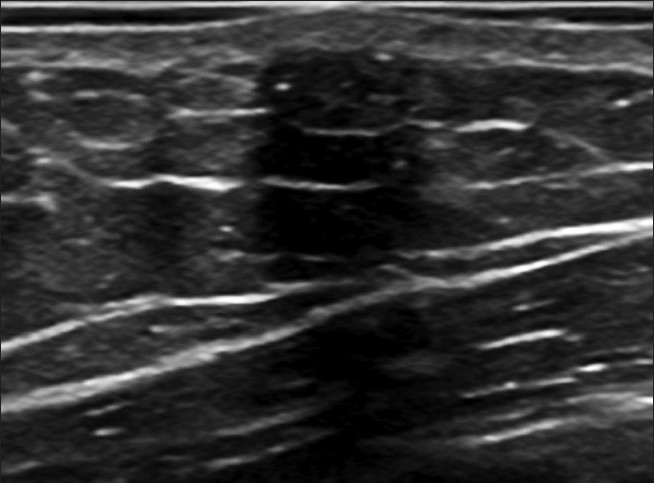
A gray-scale ultrasound image of the lesion shows a well-defined isoechoic oval mass, parallel with the skin measuring 7 mm × 3.5 mm, containing the small hyperechoic foci of calcification observed on the mammogram.

A gray-scale ultrasound image of the lesion shows a well-defined isoechoic oval mass, parallel with the skin measuring 7 mm × 3.5 mm, containing the small hyperechoic foci of calcification observed on the mammogram.