Figure 12.

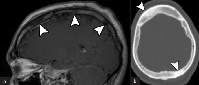
Skull Involvement. (a) Sagittal post-contrast T1-weigthed MRI shows multiple non-enhancing hypointense intraosseous skull lesions (arrowheads). (b) The corresponding axial CT image shows that the lesions are sclerotic (arrowheads).

Skull Involvement. (a) Sagittal post-contrast T1-weigthed MRI shows multiple non-enhancing hypointense intraosseous skull lesions (arrowheads). (b) The corresponding axial CT image shows that the lesions are sclerotic (arrowheads).