Figure 8.

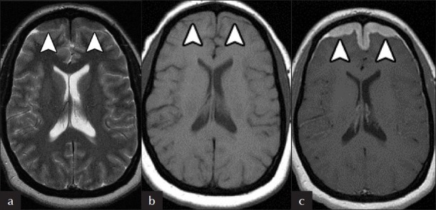
Dural Involvement. There is (a) hypointense T2 and (b) isointense T1signal in the (c) symmetrically thickened frontal lobe dura, which avidly enhance on post-contrast T1 MRI (arrowheads).

Dural Involvement. There is (a) hypointense T2 and (b) isointense T1signal in the (c) symmetrically thickened frontal lobe dura, which avidly enhance on post-contrast T1 MRI (arrowheads).