Figure 3.
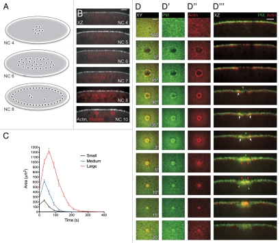
Single cell wound repair in the early Drosophila embryo. (A) Cartoon of the early Drosophila embryo (Nuclear cycle 4 to 8; NC). The early embryo is a syncytium wherein the nuclei divide in the interior of the embryo without cytokinesis through nuclear cycle 8, thereby forming a large multinucleate cell. (B) Orthogonal view of embryos expressing actin and nuclear markers. Nuclei remain away from the cell periphery until NC10, allowing for the study of wound repair before this time point without the complication of damage to the nuclei. (C) Wound repair curves (area vs. time) of small, medium and large wounds follow a stereotyped response composed of three steps and independent of wound size. Post wounding the area expands, followed by a period of rapid contraction of wound area, and then a slower phase of closure as the wounded area is remodeled. (D–D‴) Time-lapse series following wound repair in embryos expressing plasma membrane and actin markers. Plasma membrane is recruited from the area surrounding the wound and by the trafficking of vesicles to the wound site. By 90″ post wounding, the membrane has formed a plug over the wounded area. Actin is recruited from around the wound and accumulates to form a tight ring around the plasma membrane plug, which progressively contracts until the ring closes 5′ post wounding. Scale bars: XY are 20 µm and XZ 10 µm unless otherwise indicated. (Figure reprinted with permission from Abreu-Blanco et al.; J Cell Biol 2011; DOI:10.1083/jcb.201011018).29

