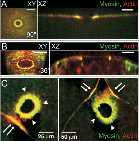
Figure 5.
Relationship between single cell and multicellular wound repair. (A) Actin and Myosin II accumulate in a ring surrounding the wound in the early Drosophila embryo. Surface projection and orthogonal view of actin and myosin II show both proteins co-localizing to form an actomyosin ring. [©Abreu-Blanco et al. Originally published in Journal of Cell Biology 2011; DOI: 10.1083/jcb.201011018].29 (B) An actomyosin purse string is observed at the leading edge of multicellular wounds in late Drosophila embryos. Surface projection and orthogonal view of actin and myosin II show both proteins co-localizing to form an actomyosin purse string. (C) Actin and myosin II accumulate at the leading edge of the wound edge as well as at cell junctions near the wound in cellularized Xenopus embryos. This accumulation at the cellular junction is distance dependant and only junctions proximal to the wound will form these secondary accumulations. (Scale bars: XY are 20 µm and XZ 10 µm unless otherwise indicated). (Figure adapted and reprinted from Clark. Curr Biol 2009; 19:1389–95 with permission from Elsevier.)32

