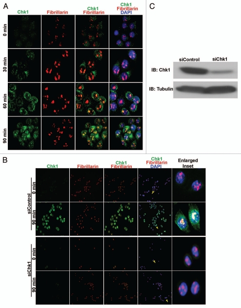
Figure 1.
Chk1 translocates to the nucleoli in response to irradiation. (A) Merged images at 40× magnification of irradiated HeLa cells stained with Chk1 (green), fibrillarin (red) and DNA (DAPI-blue) show translocated Chk1 foci in the nucleoli and colocalization with fibrillarin in a time-dependent manner from 0–90 min. (B) Merged images at 20× magnification of irradiated HeLa cells stained with Chk1 (green), fibrillarin (red) and DNA (DAPI-blue) show Chk1 colocalized with fibrillarin at 90 min in siControl-treated HeLa cells and not in siChk1-treated HeLa cells. Enlarged inset shows colocalization of Chk1 with fibrillarin (yellow arrows) at 90 min in irradiated siControl HeLa cells and not in irradiated siChk1 HeLa cells (yellow arrows). (C) Immunoblotting (IB) with Chk1 antibody confirms reduction of Chk1 levels in siChk1-transfected HeLa cells as compared to siControl-transfected HeLa cells.

