Figure 6.
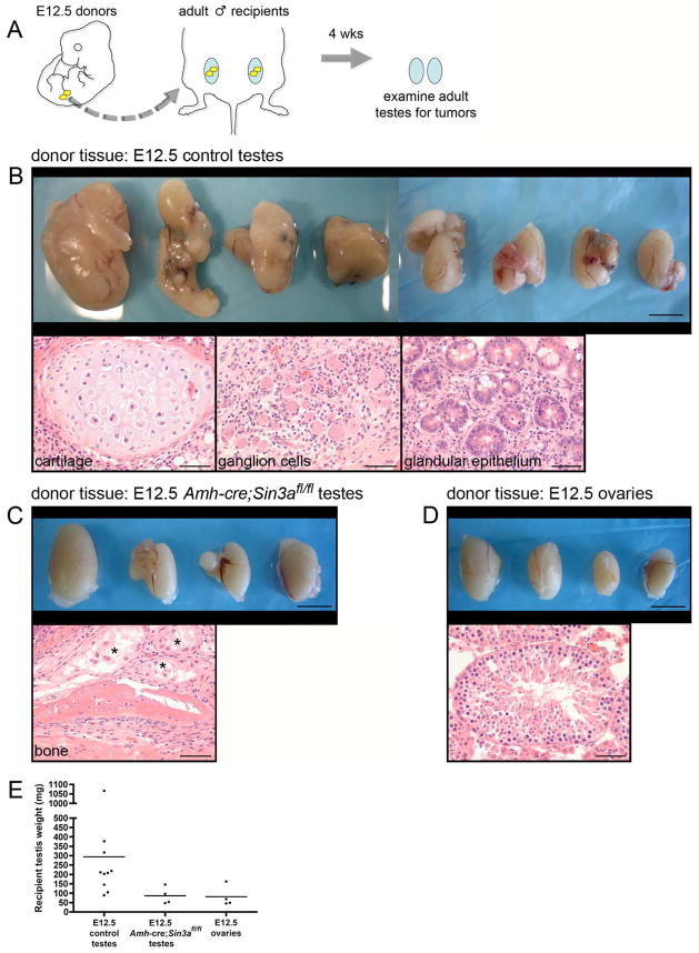
Transplanted fetal Amh-cre;Sin3afl/fl testes suppress the formation of adult testicular germ cell tumors.
(A): Schematic diagram of the transplantation assay. Gonads from E12.5 embryos (yellow) were isolated and transplanted to the testicular parenchyma of age-matched adults (blue). Four weeks later, adult testes were removed and examined for teratomas. (B, top panel): Adult testes that received E12.5 control testes as donor tissue. Testes are irregularly enlarged, containing multilobular tumors. (B, bottom panels): Tumors from all of the testes are comprised of well-differentiated tissue arising from all three embryonic germ layers (mesoderm, endoderm, neuroectoderm), consistent with teratomas. (C, top panel): Adult testes that received E12.5 Amh-cre;Sin3afl/fl testes as donor tissue, exhibiting modest enlargement. (C, bottom panel): Representative example of a teratoma from one testis containing newly formed bone. Asterisks denote degenerating seminiferous tubules. (D, top panel): Adult testes that received E12.5 ovaries as donor tissue. (D, bottom panel): Seminiferous tubule containing spermatogenic cells. (E): Weights of adult testes that received E12.5 control testes, E12.5 Amh-cre;Sin3afl/fl testes, and E12.5 ovaries as donor tissue. Scale bars represent 5 mm in the top panels (B–D), and 50μm in the bottom panels (B–D).

