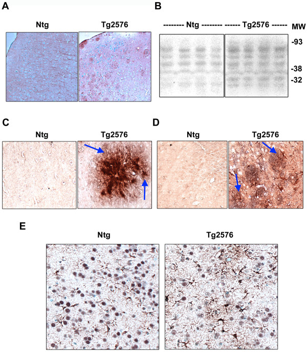
Figure 3.
Markers of oxidative stress in Alzheimer's mouse brain. (A) Brain sections of 15-17 month old nontransgenic (Ntg) and Tg2576 mice were immunostained for HNE with NovaRED and counterstained with methyl green. (B) Western blot analysis of mouse brain samples for protein bound HNE was performed. (C) Double immunostaining of mouse brain sections was carried out with monoclonal antibodies to Aβ (4G8) and polyclonal antibody for MDA. The chromogens were NovaRED for Aβ and DAB for MDA. (D) Double immunostaining of mouse brain sections was carried out with monoclonal antibody to GFAP (NovaRED) and polyclonal antibody to MDA (DAB). (E) Double immunostaining was performed with monoclonal antibody to GFAP (NovaRED) and polyclonal antibody for CREB (DAB). Representative images are provided. Markers of oxidative stress were elevated in Tg2576 mouse brain.

