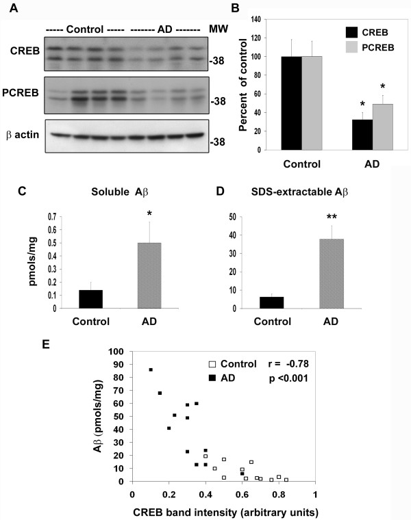
Figure 4.
Decrease in CREB protein in AD-postmortem brain. (A) Post-mortem hippocampal samples (12 each) of AD cases and age-matched controls with equal protein content were electrophoresed, transferred, and immunoprobed with antibodies to CREB, phospho CREB (PCREB) and β actin. Representative blots are shown. (B) Band intensities of CREB and PCREB were quantitated and corrected for β actin for 24 samples. Total and phospho CREB levels were significantly low in AD brain. *P < 0.01 compared to control. (C, D) Aβ 1-42 levels were determined in soluble and SDS-extractable fractions of post-mortem samples by sandwich ELISA. Elevated levels of soluble and SDS-extractible Aβ were observed in AD brain. *p < 0.01; **p < 0.001 vs control. (E) When the levels of SDS-soluble Aβ were plotted against CREB band intensities, an inverse correlation between the two parameters was observed. Control values are shown as open squares and AD values as filled squares.

