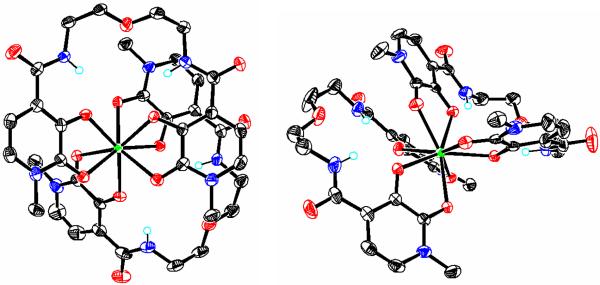
Figure 2.
X-ray crystal structures showing views of the two independent Yb(1) (left) and Yb(2) (right) complexes found in the asymmetric unit for the [Yb(5LIO-Me-3,2-HOPO)2] complex. Co-crystallized solvent molecules, and selected H atoms have been omitted for clarity and non-H atoms are drawn at the 50 % probability level (C = black, O = red, N = blue, Yb = green). Only the major component of the disordered ligand backbone is shown for the Yb(2) complex. The position of the proton necessary for charge neutrality could not be located from the difference map (see text).

