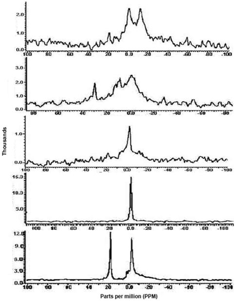
Figure 14. [31P]NMR spectra of purified IIGlc preparations of pellet (Pt), initial, middle and tail parts of peak 1 (P1-A, P1-B and P1-C, respectively) and peak 2 (P2) of E. coli strain BW25113ΔptsGΔmalE::km(pMALE-ptsG).
The spectra were recorded at 37°C, 46,000 scans and at an acquisition time of 1.6 seconds. Peak 2 was prepared from cells subjected to in vivo cross linking. For details see Materials and Methods. The spectra of these tested preparations when examined at 25, 37 and 45°C and at different scans were shown in Figs. S1 to S5 (Saier's web site).

