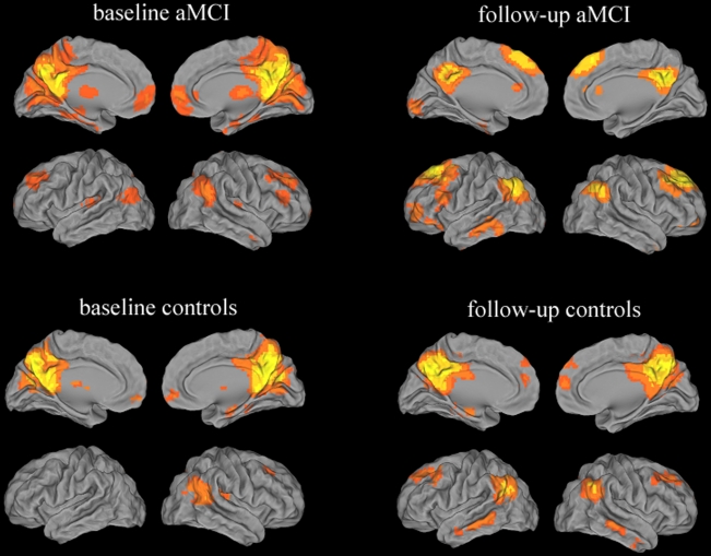
Figure 1. Validation of the ICA approach in aMCI subjects and healthy controls.
Images showed the DMN in aMCI group and controls group at baseline and follow up, separately. Significant consistency between these studies is demonstrated across the majority of clusters including the posterior cingulated cortex, precuneus, inferior parietal lobule, prefrontal cortex, ventral anterior cingulate cortex, lateral temporal cortex. Thresholds were set at a corrected P<0.05, determined by Monte Carlo simulation.

