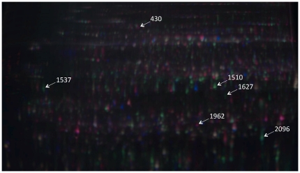
Figure 2. 2-D DIGE master gel overlay.
Gel image (pI 4–7) showing proteins derived from Manchurian ash (Cy3 – red spots) and black ash (Cy5 – green spots). The internal standard (composed of equal parts from all ash protein extracts) is displayed as blue spots. Yellow spots are those common to all species. Numbered spots identify putative defense and susceptibility related proteins (see Tables 2 and 3).

