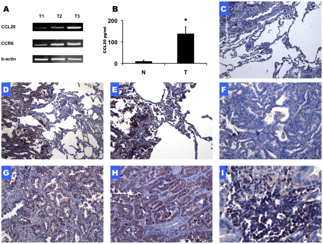
Figure 1. Expression of CCR6/CCL20 in NSCLC tissue samples.
PCR signal for CCL20, CCR6 and beta-actin in three NSCLC tumor samples (A). CCL20 protein levels in NSCLC tumors and in tumor adjacent lung tissue (n = 3) (B). Representative immunohistochemistry staining for CCL20 and CCR6 in lung adenocarcinoma tissue sections. CCL20 - Low X10 (D) and high power X20 (E) magnification. CCR6 - Low power (X10) (F, G) and high power (X20) (H) magnification. Immune cell infiltrates located with in lung adenocarcinoma tumor stain positive for CCR6 (X40) (I). Negative control staining (X10) is shown (C). (*P<0.05)

