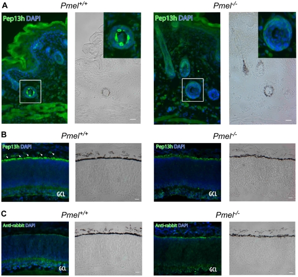
Figure 4. Immunohistochemistry of skin and retina from Pmel+/+and Pmel−/−mice.
Sections of skin (A) or retina (B, C) from Pmel+/+ (a, b) and Pmel −/− (c, d) mice were immunolabeled with αPep13h (anti-PMEL) (A, B) or secondary antibody alone (C) and counterstained with DAPI to label nuclei, and analyzed by immunofluorescence microscopy (a, c) or bright field microscopy to assess pigmentation (b, d). (A) PMEL-immunoreactive cells were found in the hair follicles of the skin in wild-type mice (a–b), but not in the Pmel −/− mice (c–d). Insets, two-fold enlargement of the boxed region. (B) αPep13h immunoreactive cells (green) were found in the choroid (arrowheads) and in the RPE from the Pmel+/+ mouse (a), but only in the RPE/ Bruch's membrane from the Pmel −/− mouse (c). GCL, ganglion cell layer. (C) Immunostaining obtained using only the secondary anti-rabbit antibody shows a signal from Bruch's membrane. Size bar, 20 µm.

