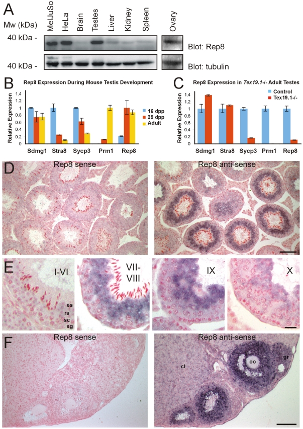
Figure 4. Expression of Rep8.
(A) The indicated rat tissues and cell extracts were analyzed by SDS-PAGE and blotting using antibodies specific for Rep8 (upper panel) and tubulin (lower panel). Rep8 was expressed almost exclusively in testes, ovaries and in the cell types used in this study. (B) qRT-PCR for Rep8 expression during mouse testis development. Bars indicate mean expression relative to β-actin, normalized to the maximum expression level for that gene during the developmental time course. Error bars indicate standard errors. (C) qRT-PCR for Rep8 expression in adult Tex19.1−/− testes. Mean expression levels relative to β-actin were normalized to control adult testes for each gene. Error bars indicate standard errors. (D) In situ hybridization of Rep8 to adult mouse testes. Bound sense or anti-sense Rep8 probes were visualized with dark blue/purple precipitate. Sections were counterstained with nuclear fast red. Scale bar 100 µm. (E) Higher magnification images of Rep8 in situ hybridization to adult testes. The approximate seminiferous epithelial stage is indicated by roman numerals, and examples of mitotic spermatogonia (sg), meiotic spermatocytes (sc), round spermatids (rs) and elongated spermatids (es) are annotated. Scale bar 20 µm. (F) In situ hybridization of Rep8 to adult mouse ovary. Scale bar 100 µm. Granulosa cells (gr), oocytes (oo) and corpora lutea (cl) are indicated.

