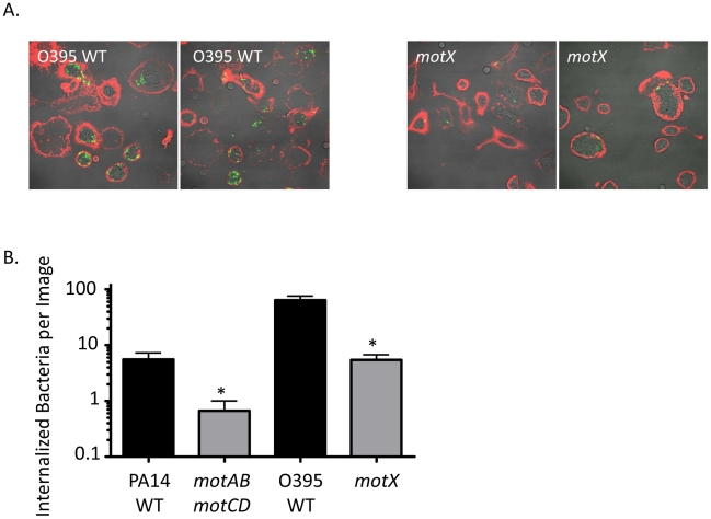
Figure 2. Fluorescence microscopy of phagocytic interactions with GFP-expressing bacteria.
(A) Confocal fluorescence microscopy of untreated murine peritoneal macrophages co-incubated at 37°C for 45 minutes with GFP-transformed V. cholerae O395 WT (left) or motX (right), washed, and subsequently stained on ice with Alexa647-conjugated wheat germ agglutinin (WGA). (B) Internalized bacteria, as in (A), were quantified on the basis of being within a contiguous WGA-decorated phagocyte plasma membrane and not co-localizing with WGA (co-localization seen as yellow, as at the plasma membrane or being external to a phagocytic cell). N≥6 images, *p<0.05.

