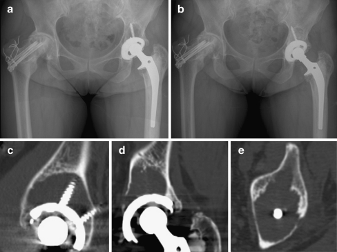
Fig. 4.
A 48-year-old woman who underwent total hip arthroplasty in the left hip joint. a Postoperative 14.7-year radiograph of the hip in the same patient. Harris hip score was 59 points in the left side. b Liner exchange with bone graft was performed c–e Selected images (sagittal, coronal, and axial) from the computed tomography scan showing increased areas of osteolysis around the acetabular component compared to Fig. 1. c–e Total pelvic osteolytic volume was 22 cm3

