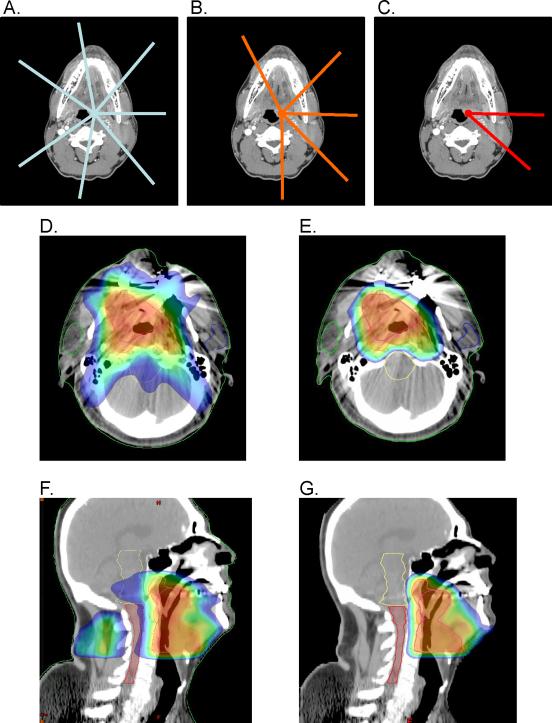
Figure 1.
Beam arrangements and treatment planning images. Representative beam arrangements used for A) photon plans to treat PTV70 with a seven-field technique, B) proton plans to treat PTV50 and PTV60 with a five-field technique, and C) proton plans to treat PTV70 with a two-field technique. Beam angles for photon plans were equally spaced and centered on each corresponding PTV. Beam angles for proton plans were individualized for each patient to minimize dose to critical structure and maintain optimal PTV coverage and dose homogeneity throughout the target volumes. Representative treatment planning images for a patient with cT4N2cM0 stage IVA squamous cell carcinoma of the oropharynx (base of tongue) in axial planes for D) IMRT and E) proton therapy, and in sagittal planes for F) IMRT and G) proton therapy. Images depict treatment to PTV70, the final treatment conedown targeting gross disease with margin. The same slices from the same initial CT data set were employed for Figures 1D and 1E and Figures 1F and 1G. Color coding: red = 100% to blue = 50% of 6Gy in 2Gy fractions.

