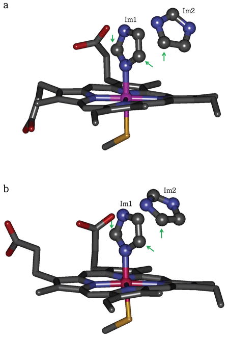
Figure 1.
Heme active sites in murine iNOS heme domain structures (a) 1NOS and (b) 1NOC, which are the only available structures of imidazole-bound iNOS. There are two nearby imidazole molecules in the active site: Im1 (coordinated to the heme iron) and Im2 (H-bonded to Glu371). Note that the positions of Im2 are different in the two structures. The arrows point at the carbon atoms whose bound hydrogens (D2 and D5 of Im1 and D5 of Im2) make the largest contributions to the observed ENDOR spectra in this study.

