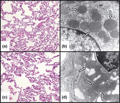
Figure 3.

Representative light (LM) and electron (EM) photomicrographs, respectively, of control (a and b) and E. coli-treated (c and d) feline lung tissue. No significant difference was observed in the histological appearance of the alveolar epithelium or the epithelial mitochondrial ultrastructure between the control and E. coli-treated animals after 6 h. In addition, the mitochondria (white arrows) appeared intact and did not show the typical features of cytopathic injury, namely swelling and loss of cristae. (LM – staining: haematoxylin and eosin; original magnification: 20×, EM – staining: uranyl acetate and lead citrate; original magnification: 20,000×).
