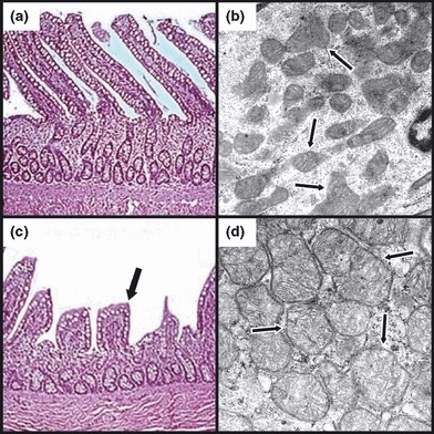
Figure 4.

Representative light (LM) and electron (EM) photomicrographs, respectively, of control (a and b) and Escherichia coli-treated (c and d) feline ileal tissue. Escherichia coli-treated specimens (c) demonstrated retraction (black arrow) and occasional loss of villi compared to matching controls (a). In contrast to observations in the lungs, dramatic changes in mitochondrial morphology were evident in the ileum following E. coli treatment (d), evidenced by mitochondrial swelling with a loss of well-defined cristae (black arrows). (LM – staining: haematoxylin and eosin; original magnification: 20×, EM – staining: uranyl acetate and lead citrate; original magnification: 55,000×).
