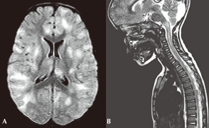
Fig. 2.
(A) An axial fluid-attenuated inversion recovery magnetic resonance imaging (MRI) of the brain in a child with acute disseminated encephalopmyelitis demonstrates multifocal areas of hyperintensity in both cerebral hemispheres involving cortical gray matter, centrum semiovale, and deep gray nuclei. (B) A saggital T2-weighted MRI of the spine in the same child demonstrates high signal intrinsic to the spinal cord, consistent with longitudinally extensive transverse myelitis.

