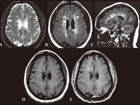
Fig. 3.
Axial T2-weighted (A) and axial fluid-attenuated inversion recovery (FLAIR) (B) images show multiple, ovoid shaped, hyperintense foci in the periventricular area, consistent with multiple sclerosis plaques. Sagittal FLAIR (C) image also shows these lesions to be radiating out from the corpus callosum. Axial precontrast T1-weighted (D) image shows that many of these lesions are hypointense, consistent with black holes. Axial postgadolinium fat saturated T1-weighted (E) image shows that some of these plaques enhance in a ring-like fashion consistent with active plaques.

