
Figure 5.
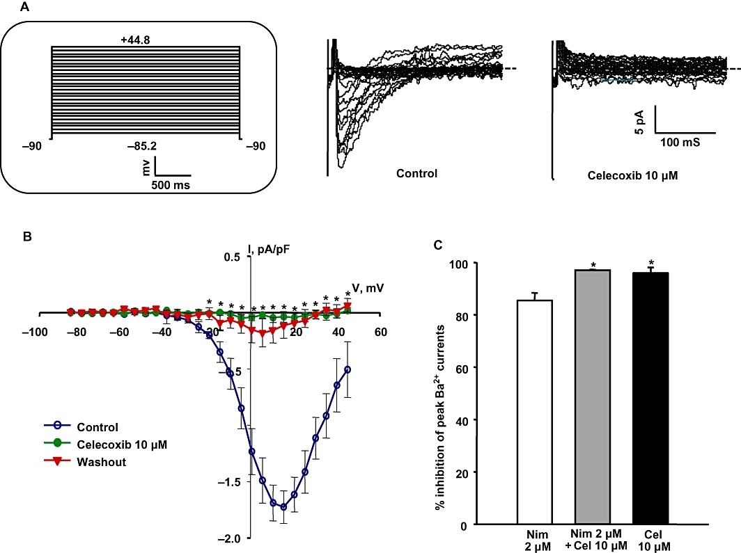
Celecoxib inhibits voltage-sensitive calcium currents in basilar artery myocytes. (A) Representative voltage-sensitive Ba2+ current (IBa) traces before and during the application of 10 µM celecoxib. Inset shows the schematic of the voltage protocol used to record IBa. (B) Summarized I-V curves show the effect of 10 µM celecoxib on IBa. Celecoxib significantly inhibited IBa at all tested voltages from −20.2 to +39.8 mV (n = 5, *P < 0.05, Student's t-test). (C) Percentage inhibition of peak IBa with the application of 2 µM nimodipine alone, 2 µM nimodipine + 10 µM celecoxib and 10 µM celecoxib alone. Celecoxib either alone or when added along with nimodipine produced significantly more inhibition of IBa compared with nimodipine alone (n = 5–6, *P < 0.05 using one-way anova followed by post hoc Holm-Sidak test).