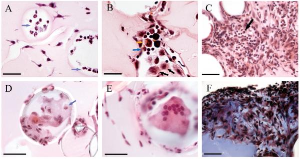
Fig. 9. Cells associated with BCG lipid-coated beads.
Matrices containing BMMØ and BCG lipid-coated beads were injected i.p. and recovered at 14 h (A), 4 days (B) and 12 days (C - F). (A): Neutrophils (blue arrows) were present in spaces that had been occupied by beads after 14 hr. Scale bar, 35 mm. (B): A diverse infiltrate of macrophages (black arrows), neutrophils (blue arrow), eosinophils (orange arrow) and lymphocytes (small cells with little cytoplasm) accumulated at beads at 4 days. Scale bar, 25 mm. (C): A dense cellular infiltrate composed of mononuclear and polymorphonuclear leukocytes and lymphocytes, including plasma cells (black arrow) formed at 12 days. Infiltrates were especially florid in spaces between lipid-coated beads. Scale bar, 35 mm. (D): Epithelioid macrophages and occasional neutrophils (blue arrow) typically adhered to beads. Scale bar represents 50 mm. (E): Multi-nucleated giant cell in association with a bead at 12 days. Scale bar, 35 mm. (F): Fibrotic material (dark blue-staining material) was deposited in the dense aggregates of leukocytes at 12 days. The uniformly staining blue material in the lower left corner of the panel is the collagen in the Matrigel. Scale bar, 35 mm. Representative H & E-stained sections (A – E) and a trichrome-stained section (F) are shown. Reproduced from (97).

