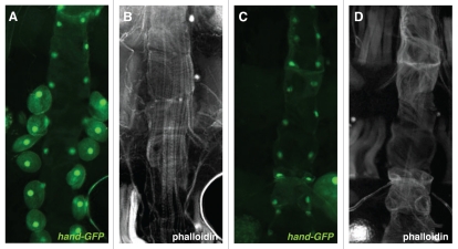
Figure 4.
Visualization of the adult heart. (A and B) Control cross: hand-GFP; hand-Gal4 with y1w67c23. (C and D) hand-GFP; hand-Gal4 crossed with UAS-M2(H37A)-3ME. Dorsal vessel myofibrils of dissected adult females (≤2 days old) were visualized with Alexa594-conjugated phalloidin, with cardioblasts and pericardial cells marked by hand-GFP. All pericardial cells except for one, in one third of population screened, were ablated by crosses with the M2(H37A) toxin. As a result of the ablation crosses, all dorsal vessels were characterized by abnormally organized myofibrils but maintained cardioblast GFP expression.

