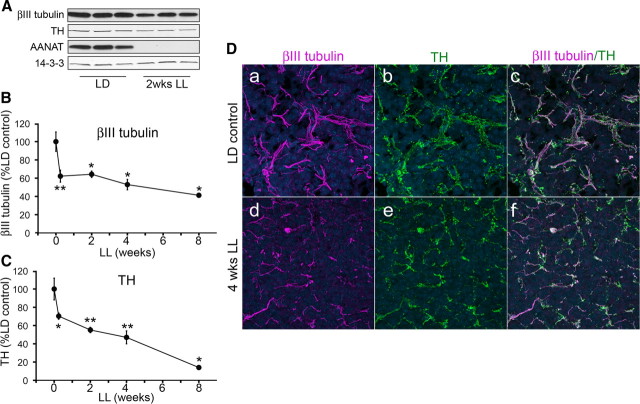
Figure 1.
Constant light results in a progressive decrease in expression of axonal proteins in the pineal. A, Representative Western blot analysis of pineal lysates from animals reared in either LD conditions or following 2 weeks of LL, probed with antibodies against βIII tubulin, TH, AANAT, and 14-3-3. In this and all other Western blots, each lane represents pineal lysate from a different animal and expression of 14-3-3 is monitored for quantifying protein levels. B, βIII tubulin protein levels following light exposures of increasing durations (0, 0.25, 2, 4, and 8 weeks). The values are expressed as percentage relative to LD control animals, which are as follows: 62% at 0.25 weeks (42 h; n = 6), 64% at 2 weeks (n = 3), 53% at 4 weeks (n = 3), and 41% at 8 weeks of LL (n = 3). C, TH expression following exposure to constant light is 70% (n = 6) after 0.25 weeks, 55% (n = 3) after 2 weeks, 47% (n = 3) after 4 weeks, and 14% (n = 3) after 8 weeks. Two-tailed t test was performed for statistical analysis; *p < 0.05, **p < 0.01. Error bars represent SEM. D, Pineal glands from control animals reared in LD conditions (a–c) and after 4 weeks of LL (d–f) were analyzed by immunohistochemistry for the presence of axonal markers βIII tubulin (a, c, d, f) and TH (b, c, e, f). Note colocalization of βIII tubulin and TH and marked decrease in the thickness and density of axonal terminal arbors (d–f) following exposure to constant light. Hoechst 3332 was used as nuclear counterstain (blue). All animals were killed at ZT18. The findings shown in D were confirmed in two rats. AANAT levels were assayed in all Western blotting experiments to ascertain the silencing of adrenergic activities when light was present at night.

