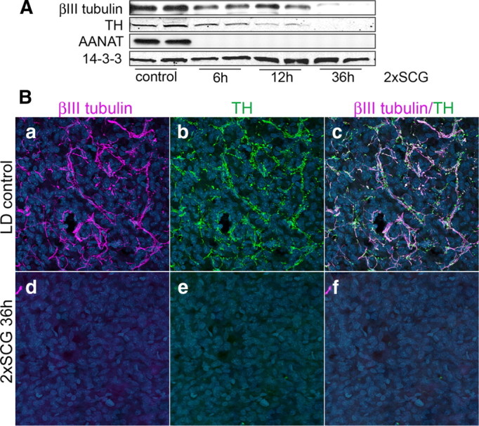
Figure 2.

Axonal proteins in the pineal represent mainly sympathetic axonal arbors originating from the two SCGs. A, Western blot analysis of βIII tubulin and TH in pineals from control and ganglionectomized animals (2×SCG) 6, 12, and 36 h after surgical denervation. Note the progressive decrease in TH (64% of control at 6 h, 17% at 12 h, and 1% at 36 h 2×SCG) and βIII tubulin (53% of control at 6 h, 40% at 12 h, and 3% at 36 h postdenervation) proteins following bilateral ganglionectomy. Expression of AANAT, which is induced by norepinephrine release from the SCG axon terminals, was monitored to confirm the surgical denervation. B, Pineal glands from control (a–c) and 36 h following 2×SCG (d–f) were analyzed by immunohistochemistry for the expression of axonal markers βIII tubulin (a, c, d, f) and TH (b, c, e, f). Hoechst 3332 was used as nuclear counterstain (blue). Note that βIII tubulin and TH colocalize in axonal fibers in the control sample (a–c) and that, 36 h following surgical denervation, axonal proteins are undetectable. All animals were killed at ZT15. These results (A, B) were confirmed in >5 independent experiments.
