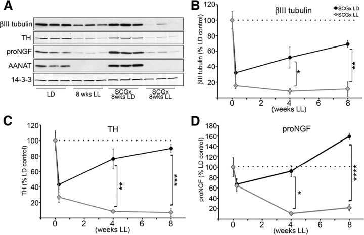
Figure 5.
Axonal growth from uninjured SCG neurons following unilateral SCG removal is profoundly impaired in the absence of neural activity. A, A representative Western blot of pineal lysates from control animals kept in LD or LL, and from animals which had one SCG surgically removed, and were maintained after surgery for 8 weeks in LD (SCGx 8wks LD) or in LL (SCGx 8wks LL). Membranes were probed with antibodies against βIII tubulin, TH, NGF, AANAT, and 14-3-3. B, Time course of βIII tubulin levels in the pineal following unilateral ganglionectomy (0, 0.25, 4, and 8 weeks) from animals reared in either LD (SCGx LD) or LL (SCGx LL). Values are expressed as percentage relative to LD control animals: 32% at 0.25 weeks (42 h; n = 6), 52% at 4 weeks (n = 3), and 69% at 8 weeks (n = 3) in the LD samples; and 14% at 0.25 weeks (n = 6), 8% at 4 weeks (n = 3), and 11% at 8 weeks (n = 3) in animals maintained in LL conditions. C, Time course of TH protein following unilateral ganglionectomy from animals reared in either LD or LL. Values are relative to LD controls: 43% at 0.25 weeks (42 h; n = 6), 76% at 4 weeks (n = 3), and 90% at 8 weeks (n = 3) in the LD samples; and 27% at 0.25 weeks (n = 6), 8% at 4 weeks (n = 3), and 7% at 8 weeks (n = 3) in LL samples. D, Time course of proNGF (∼34 kDa) expression following unilateral ganglionectomy from animals reared in either LD or LL, expressed as percentage relative to LD control animals: 67% at 0.25 weeks (42 h; n = 6), 92% at 4 weeks (n = 3), and 159% at 8 weeks (n = 3) in the LD samples; and 64% at 0.25 weeks (n = 6), 11% at 4 weeks (n = 3), and 22% at 8 weeks (n = 3) in animals maintained in LL conditions. Unpaired, two-tailed t test was performed for statistical analysis comparing the time point values for each protein to the LD control values. The values between the SCGx LD and SCGx LL at each time point: *p < 0.05, **p < 0.01, ***p < 0.001, ****p < 0.0001. Error bars represent SEM. All animals were killed at ZT18. Again, silencing of the sympathetic activity by light was confirmed by the absence of AANAT protein in the pineal gland of rats exposed to light at night. Of note, histological confirmation of sprouting following partial pineal denervation has been previously demonstrated (Lingappa and Zigmond, 1987).

