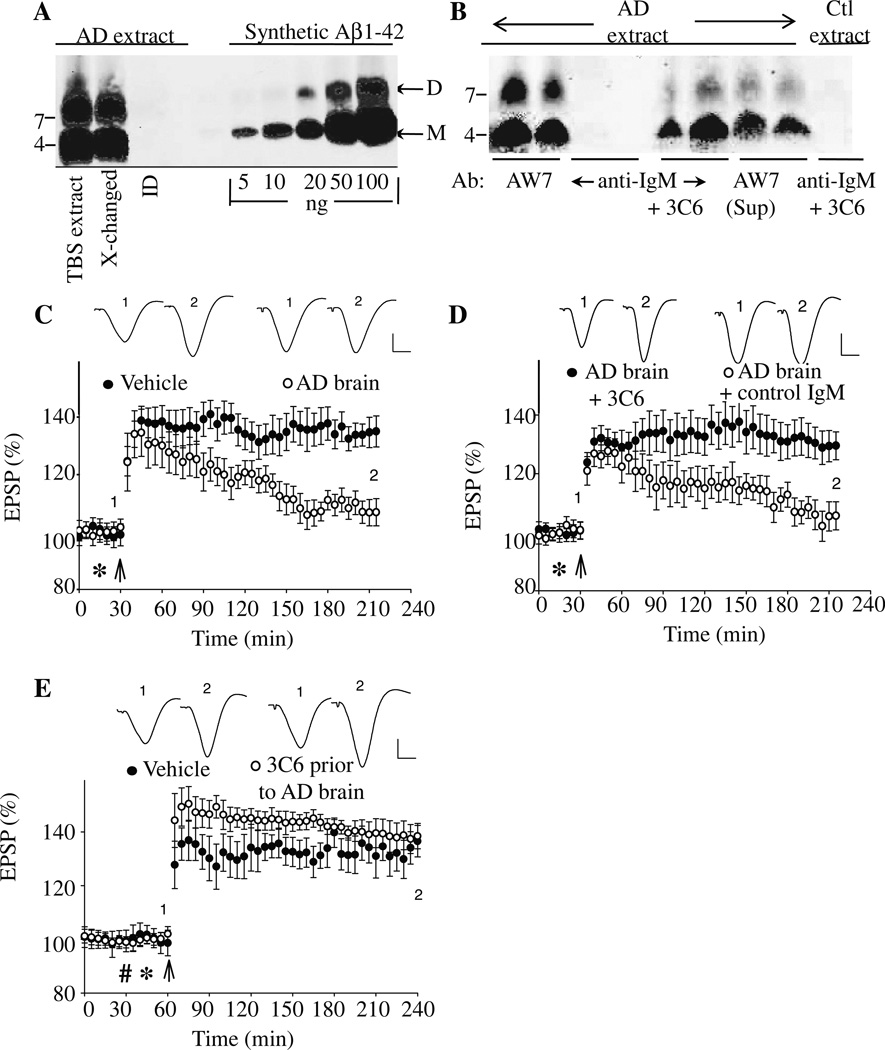
Figure 4. MAb 3C6 binds Aβ and ameliorates the block of LTP induced by AD brain extract.
A TBS extract of AD brain was examined by immunoprecipitation (IP)/Western blotting using the polyclonal anti-Aβ antibody, AW8, for IP and a combination of anti-Aβ mAbs, 2G3 and 21F12, for Western blotting (A). The first two lanes of the Western blot show that the untreated TBS extract and buffer-exchanged extract contained highly similar amounts of Aβ monomer and SDS-stable Aβ dimers. The third lane shows that the first round of IP had effectively depleted the extract of all detectable Aβ. Molecular weight standards are indicated on the left, and Aβ monomers (M) and dimers (D) labeled with arrows on the right. Based on standard synthetic Aβ1-42 included on the blot, the test samples contained 50 ng/ml and 70 ng/ml Aβ dimer and monomer, respectively (B). Western blot of Aβ IP’d from AD brain TBS extract by the anti-Aβ polyclonal IgG, AW7; anti-IgM alone, and mAb 3C6 are shown in duplicate in lanes 1–6. To determine if 3C6 bound all the Aβ present in the AD TBS extract, the material not IP’d by 3C6 was used for IP with AW7 (lanes 7+8). Similarly, to determine the specificity of the bands detected by 3C6, this antibody was also used to IP a TBS extract from a non-demented control (lane 9). The blot shows that both 3C6 and AW7, but not the control IgM, IP’d Aβ assemblies that migrated on polyacrylamide gels as monomers and SDS-stable dimers. (C) Intracerebroventricular (i.c.v.) injection (*) of AD TBS brain extract inhibited high frequency stimulation (HFS, arrow) induced LTP (107 ± 5 %, n = 4, baseline, p>0.05 compared with pre-HFS baseline, and p<0.05 compared with vehicle injected controls (135 ± 5 %, n = 8)). (D) Co-injection of 5 µg of 3C6 prevented the inhibition of LTP by AD TBS extract (130 ± 5 %, n = 5, compared p<0.05 compared with AD TBS alone, n = 5), whereas co-injection of a control IgMκ, M1520, did not (105± 5 %, p<0.05, n = 5, compared with AD TBS extract + 3C6, p>0.05 compared with AD TBS extract alone). (E) LTP was not impaired when 10 µg of 3C6 (hash symbol) was i.c.v. injected 15 min before AD TBS brain extract (138 ± 5 % (n= 4) baseline, p<0.05 compared with 136 ± 6 % (n = 5) vehicle injected controls). Insets show representative EPSP traces at the times indicated. Calibration bars: vertical, 1 mV; horizontal, 10 ms.

