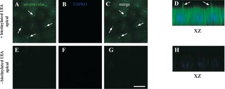
Fig. 3.
Localization of H antigen-bearing glycoproteins in polarized MDCK cells. Polarized MDCK cells were fixed, permeabilized, and incubated with or without biotinylated Ulex europeaus agglutinin (UEA). Cells were incubated with FITC-conjugated streptavidin (green), fixed, stained with TOPRO-3 (blue), and mounted for confocal microscopy. Representative images are shown for the apical pole of MDCK cells incubated with (A–D) or without (E–H) UEA are shown. Merged images (C and G) and XZ sections (D and H) are presented. Arrows indicate the localization of UEA on the primary cilia. Scale bar = 10 μm.

