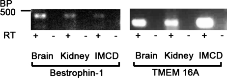
Fig. 4.
Expression of bestrophin-1 and TMEM16A mRNA in mIMCD-K2 cells. Photograph of gel showing PCR amplification products in mouse brain and kidney tissue (positive controls) and mIMCD-K2 cells (IMCD) after reverse transcription of mouse bestrophin-1 and TMEM16A mRNA. Samples containing reverse transcriptase (RT; +) or negative controls lacking RT (−) are included for each tissue/primer combination. BP 500 indicates a marker of 500 base pairs.

