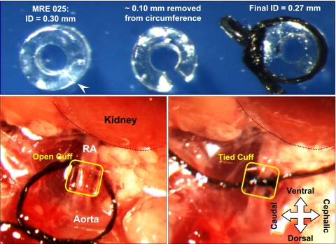
Fig. 1.
Photographs showing the use of polyurethane tubing as a cuff to constrict the renal artery. Top, left: short length (∼0.6 mm) of MRE25 tubing [inner diameter (ID) 0.30 mm] that has been sliced lengthwise with a scalpel (at arrowhead); middle: a ∼0.1-mm wedge has been removed from the wall, so that when the tubing circumference is closed using a suture, the ID is ∼0.27 mm (right). The size of the wedge can be adjusted to finely tune the final ID. Bottom: cuff is shown after it is positioned around the left renal artery (RA; left) and after the suture is tied to constrict the artery (right). The position of the kidney has been outlined and labeled, and the orientation of the mouse, in a right lateral decubitus position, is indicated.

