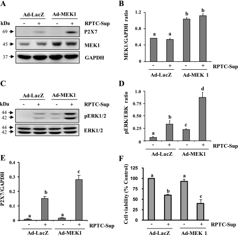
Fig. 5.
Effect of overexpression of wild-type MEK1 on P2X7 expression. NRK-49F cells were infected with Ad-Lacz or Ad-wild-type MEK1 as described in materials and methods and then treated with necrotic RPTC for 24 h (A and F) or 30 min (C). After treatment, cells were harvested and subjected to immunoblot analysis for MEK1, P2X7, p-ERK1/2, ERK1/2, or GAPDH (A and C). The levels of MEK1 (B) and P2X7 (E) were quantified by densitometry and normalized to GAPDH level. The levels of p-ERK1/2 and ERK1/2 were quantified by densitometry and the ratio between them was calculated (D). Bars with different letters (a–c) are significantly different from one another (P < 0.05). Cell viability was determined by the MTT assay (F). Values are means ± SD of 3 independent experiments conducted in triplicates and expressed as the percentage of control. Representative immunoblots from 3 experiments are shown.

