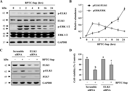
Fig. 6.
Effect of inhibition of Elk1 on necrotic RPTC-induced P2X7 expression and cell death. NRK-49F were treated with RPTC-Sup supernatant for the indicated time (0–24 h) and cell lysates were subjected to immunoblot analysis for p-Elk1, Elk1, p-ERK1/2, ERK1/2, or GAPDH (A). The phosphorylated and total levels of p-Elk1, Elk1, p-ERK1/2, and ERK 1/2 were quantified by densitometry and phosphorylated protein levels were normalized to total protein levels (B). Cultured NRK-49F cells were transfected with scrambled siRNA or siRNA specific for Elk1. At 24 h after posttransfection, cells were treated with necrotic RPTC for 24 h and cell lysates were subjected to immunoblot analysis for Elk1, pElk1, P2X7, or GAPDH (C). The cell viability was assessed by MTT (D). Values are means ± SD of 3 independent experiments conducted in triplicates and expressed as the percentage of control.

