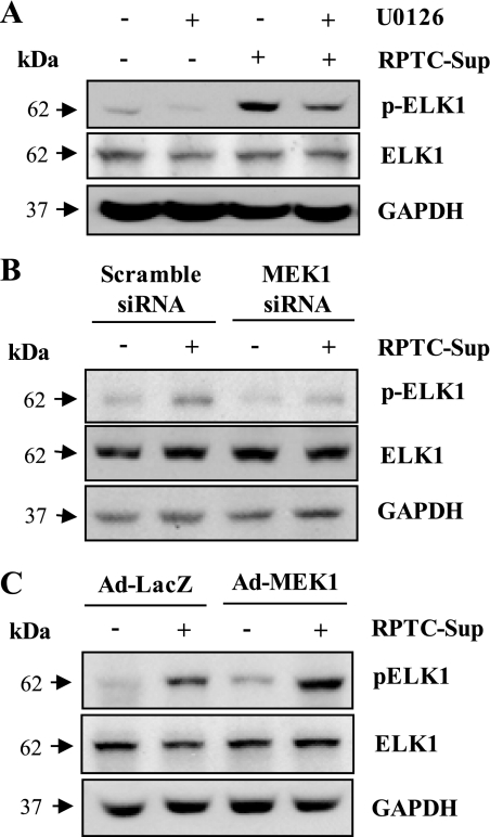
Fig. 7.
Effect of inhibition of MEK1 or overexpression of MEK1 on necrotic RPTC-induced Elk1 phosphorylation. Cultured NRK-49F cells were treated with U0126 (20 μM) for 1 h and then exposed to necrotic RPTC supernatant for 24 h (A). NRK-49F cells were transfected with siRNA specific for MEK1 (B) or infected with adenovirus encoding MEK1 (Ad-MEK1; C), and cells were treated with necrotic RPTC for 24 h. Cells were harvested and cell lysates were subjected to immunoblot analysis for pElk1, Elk1, or GAPDH. Representative immunoblots from 3 experiments are shown.

