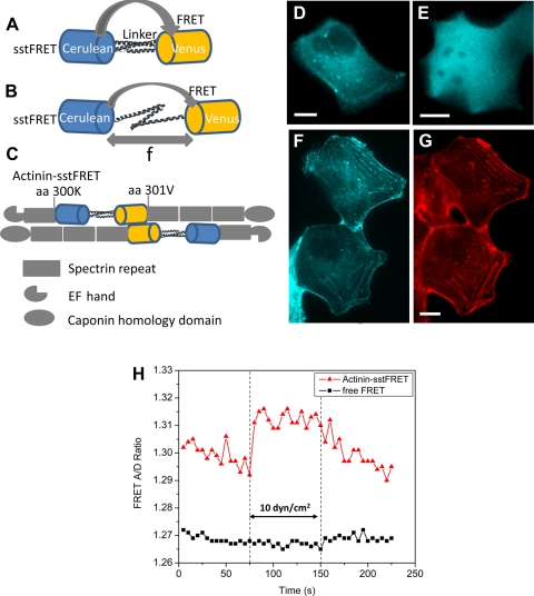
Fig. 1.
Spectrin-repeat stress-sensitive fluorescence resonance energy transfer (FRET) (sstFRET) sensor construction and calibration. A: sstFRET consists of Cerulean, donor, Venus, acceptor, and a linker. Resting sensor shows higher FRET. B: under axial force (f) the distance between donor and acceptor is extended leading to lower FRET. C: actinin-sstFRET, showing actinin hosting sstFRET at amino acid 300, close to the middle of actinin. D and E: fluorescence images [cyan fluorescent protein (CFP) channel] of Madin-Darby canine kidney (MDCK) cells expressing actinin-sstFRET and free sstFRET, respectively. F and G: fluorescence images of actinin-sstFRET in MDCK cells (F) and actin stained with phalloidin-Alexa Fluor568 (G), showing a colocalization of actinin-sstFRET with actin. H: change of FRET ratio in response to an increase of shear stress from 0.6 to 10 dyn/cm2, showing an increase of FRET in actinin-sstFRET expressing cells, not in free sstFRET expressing cells. The scale bar represents 10 μm.

