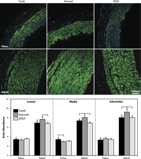
Fig. 4.
Transmural morphometry reveals an age-dependent influence of organ culture with VEGF on the regional expression of smooth muscle α-actin. Endothelium-denuded carotid arteries from fetal and adult sheep were organ cultured as described for the contractility studies and then were fixed in 4% paraformaldehyde, sectioned at 5 μm, and immunostained for smooth muscle α-actin (green signal). Cell nuclei were stained with DAPI (blue signal). For all images shown, the artery lumen faces rightward. Signal intensities were independently adjusted for each image to maximize dynamic range and do not indicate absolute marker abundance. Coronal sections were line scanned to determine the relations between fluorescent intensity and location in the artery wall. Three locations were examined in detail. The “lumen” region was defined as the area lying just inside the basal elastic lamina. The “media” region was defined as the area midway between the basal elastic lamina and the adventitial-medial border. The “adventitia” region was defined as the area of smooth muscle immediately adjacent to the adventitial-medial border. Values of fluorescent intensity recorded in each region were converted to apparent concentration using standardized calibration curves (see Fig. 3) and then averaged across multiple animals and normalized relative to Western blot measurements of smooth muscle α-actin abundance. For the Fresh segments, the letters “A” and “L” indicate significant differences compared with the adventitial and luminal regions, respectively. Asterisks indicate paired differences due to treatment (P < 0.05, ANOVA). Error bars indicate means ± SE for N = 6 for both fetal and adult arteries. Overall, smooth muscle α-actin abundance was significantly greater in adult than in fetal arteries for all corresponding treatments. Organ culture with 3 ng/ml VEGF-A165 significantly depressed smooth muscle α-actin abundance in adult but not fetal arteries. Note that organ culture altered the structure of both the medial and adventitial layers in an age-dependent manner.

