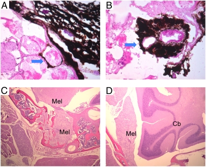
Fig. 4.
Local invasion of tail and meningeal melanomas. In the late stage of melanomagenesis, highly aggressive melanoma cells from tail or meninges penetrate the underlying muscular layer (A and B) or bone tissues (C and D). (A and B) The H&E staining of tails from TRP1-mGluR5 wild-type transgenic mice. Arrows indicate invasion of melanoma cells to skeletal muscle. (C and D) Melanin-bleached specimen followed by H&E staining of Thy1-mGluR5 S901A transgenic mice. Mel, melanoma; Cb, cerebellum.

