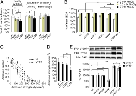
Fig. 3.
Effects of β1Y-to-F substitutions on adhesion and signaling. (A) Whereas β1 surface levels were reduced in β1Y-to-F freshly isolated keratinocytes, they became similar over time in cultured cells propagated on collagen I. (B) Quantification of 9EG7 activation epitope by FACS. (C) Spinning-disk hydrodynamic shear stress assay measuring adhesion strength. A cell detachment profile of β1YYFF and WT keratinocytes is shown as a function of applied shear stress. (D) Mean (50%) ± SD cell detachment force of four independent experiments. (E) FAK-pY397 and FAK-pY861 were assessed in keratinocytes grown on collagen-coated plastic. Results were quantified by densitometry and normalized to total FAK levels. ***P < 0.001; **P < 0.01; *P < 0.05.

