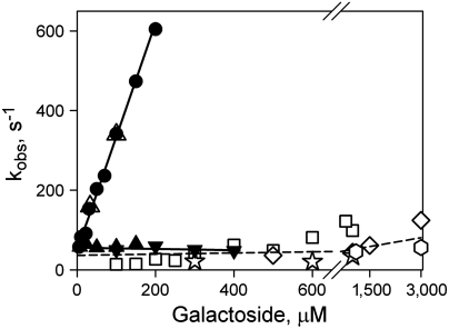
Fig. 7.
Concentration dependence of sugar-binding rates with rates of opening of the periplasmic cavity for mutant N245W/A155C. NPG binding rates measured as Trp151-NPG FRET are shown for purified mutant in DDM (●), reconstituted in proteoliposomes at LPR 10 (▴) or 5 (▾), and after addition of DDM (△). Linear fits to the data obtained for Trp151-NPG FRET are shown as solid lines. Reconstituted mutant N245W/A155C binds NPG with kobs = 56 ± 7 s-1. Rates of unquenching of Trp245 fluorescence resulting from opening of the periplasmic cavity after sugar binding are shown as open symbols: TDG (□); melibiose (⋄); octyl-α-d-galactoside (☆); methyl-α-d-galactoside (hexagon). The rate of opening of periplasmic cavity in DDM is 50–100 s-1 at the higher concentrations of the four galactosides tested.

