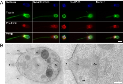
Fig. 2.
SNAREs, Munc18, and the secretory apparatus in M. brevicollis. (A) Confocal micrographs illustrate immunolabelings for syntaxin 1, synaptobrevin, SNAP-25, or Munc18, all detected at the posterior (apical) pole of the cell. (Center) A cross-section image through the posterior pole shows a circular distribution of synaptobrevin. Colabeling of tubulin and actin allow for identifying the cell's cytoskeleton/architecture. (B) Ultrastructural investigation reveals the presence of the Golgi apparatus (Go) and associated clear vesicles (ve) at the posterior pole of the cell, near the root/basal body of the flagellum (fl) (Right). Note the presence of unrelated, heterogeneous large vesicles, most probably food vacuoles (fv), at the anterior pole (Left). The cell nucleus (nu), mitochondria (mt), and the collar (co) are indicated as well. (Scale bars: A, 1 μm; B, 500 nm.)

