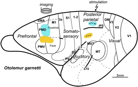
Fig. P1.
Parietal–frontal nodes in networks for reaching (blue) and face defensive (yellow) movements on a brain of prosimian primate (Otolemur garnetti). Dots in the blue region in PPC mark microelectrode penetrations where electrical stimulation evoked reaching movements and activated groups of neurons in parts of dorsal premotor (PMD) and primary motor (M1) cortex (blue oval), which was revealed by optical imaging. Stimulation of more lateral sites in the face defensive zone (yellow) of PPC evoked a grimace and closing of the eye and activated regions within a more lateral zone of frontal cortex (yellow oval), including parts of ventral premotor cortex (PMV) and the face sector of M1. The supplementary motor area (SMA), subdivisions of somatosensory cortex (3a, S1, 1–2, PV, and S2), auditory cortex, and visual areas (V1, V2, V3, DM, MT, and MST) are outlined. IPS, the intraparietal sulcus; STS, superior temporal sulcus; LS, lateral sulcus.

