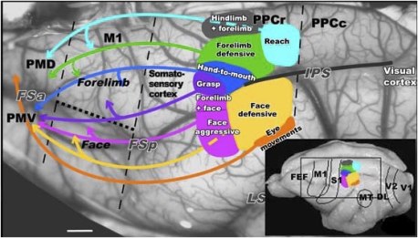
Fig. 1.
Summary of the PPC organization in galago. Functionally distinct movement zones are marked with colors on the exposed left hemisphere. Connections between functional PPC zones and frontal motor region PM-M1 are marked with color-coordinated arrows. This view matches the location of Inset on the schematic of PPC functional zones on a dorsolateral view of the hemisphere (lower right corner). Black dashed lines mark approximate borders of M1 and the border between rostral (PPCr) and caudal PPC (PPCc). The dotted line marks the border between the M1 forelimb and face representations. M1, primary motor cortex; PMD and PMV, dorsal and ventral motor areas; PPCr and PPCc, rostral and caudal posterior parietal cortex; FSa and FSp, anterior and posterior frontal sulci; IPS, intraparietal sulcus; LS, lateral sulcus; FEF, frontal eye field; DL, dorsolateral visual area; MT, middle temporal area; S1, primary somatosensory area; V1, primary visual area; V2, secondary visual area. (Scale bar: 1 mm.)

